
VISUALIZATION OF HIV
These illustrations attempt to depict HIV based on currently available structural and biochemical information. We are interested in updating the images as new information becomes available. Please feel free to send comments and critiques to goodsell@scripps.edu .
HIV-Infected Cell (David S. Goodsell 2022)
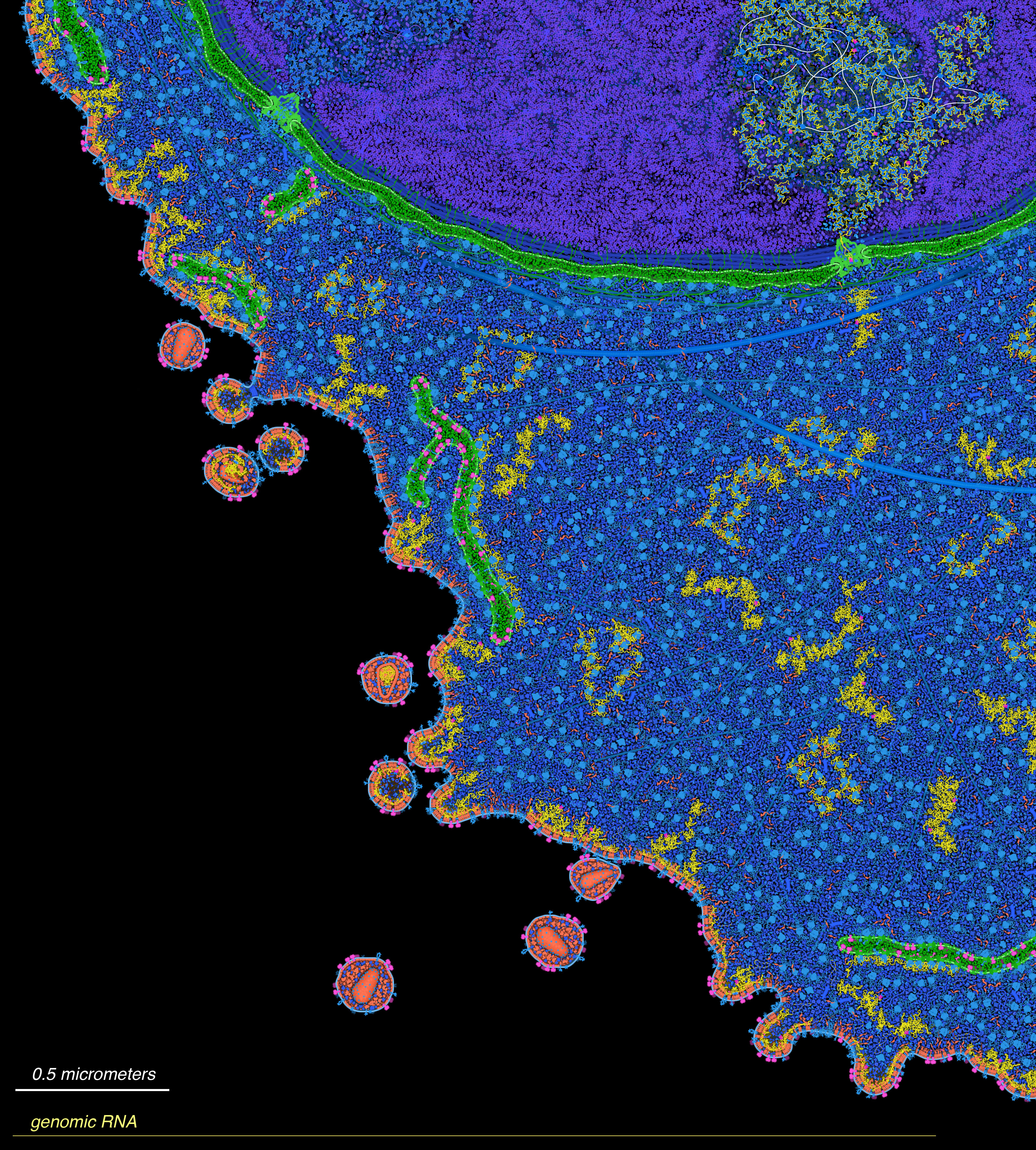
Integrative illustration of an HIV-infected cell. The cell nucleus is at the top, with DNA in purple and nuclear membrane in green. The HIV genomic DNA (thin white strand at upper right) is integrated into the cell’s genome and is being transcribed into RNA (yellow) by RNA polymerases. The RNA is then transported through the nuclear pore to the cytoplasm. Some of the RNA is translated by cytoplasmic ribosomes to build gag and gag-pol proteins (red) and by ribosomes bound to the endoplasmic reticulum (green compartments in the cytoplasm) to build envelope glycoproteins (magenta). Other copies of the genomic RNA dimerize and are packaged into virions. Finally, gag and gag-pol associate with the cell membrane, capture the dimers of genomic RNA, and bud from the surface of the cell to form new virions. Inside these virions, the proteins mature, forming the distinctive cone-shaped capsid that will deliver the genomic RNA when the virus infects a new cell. The long yellow line at the bottom represents an extended genomic RNA.
This illustration is based largely on previous illustrations of eukaryotic cells and HIV life cycle, presented in two publications:
- Goodsell, DS (2011) Eukaryotic Cell Panorama. Biochemistry and Molecular Biology Education 39, 91-101
- Goodsell, DS (2015) Illustrations of the HIV Life Cycle. Current Topics in Microbiology and Immunology 389, 243-252
This illustration is free for use. Click on the image for a full-size jpg file, or visit PDB-101 for a full-size tif file. Please use an acknowledgement such as: “Illustration by David S. Goodsell, B-HIVE Center, RCSB Protein Data Bank and Scripps Research. doi: 10.2210/rcsb_pdb/goodsell-gallery-047”
Mature and ALLINI-Inhibited HIV (David S. Goodsell 2016)
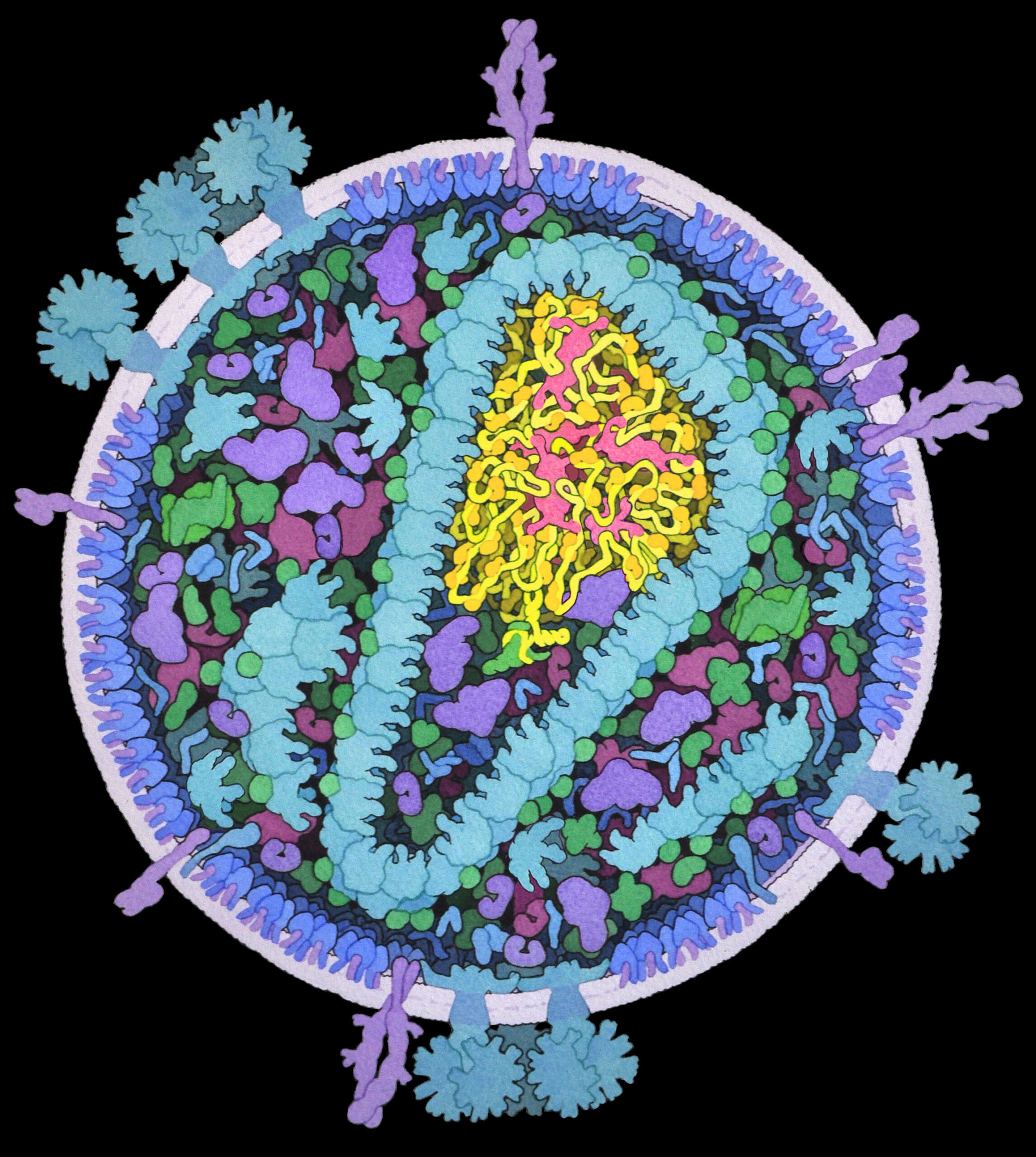
Recent results from HIVE Center researchers have revealed that integrase is important for packaging the HIV genome inside the capsid, and that ALLINIs aggregate integrase and often lead to virions with the genome outside the capsid. These two paintings show cross sections of mature HIV (left) and HIV inhibited with ALLINIs (right).
See Kessl et al. “HIV-1 integrase binds the viral RNA genome and is essential during virion morphogenesis” Cell 166, 1257-1268.
Cross section of immature HIV (2013, David S. Goodsell)
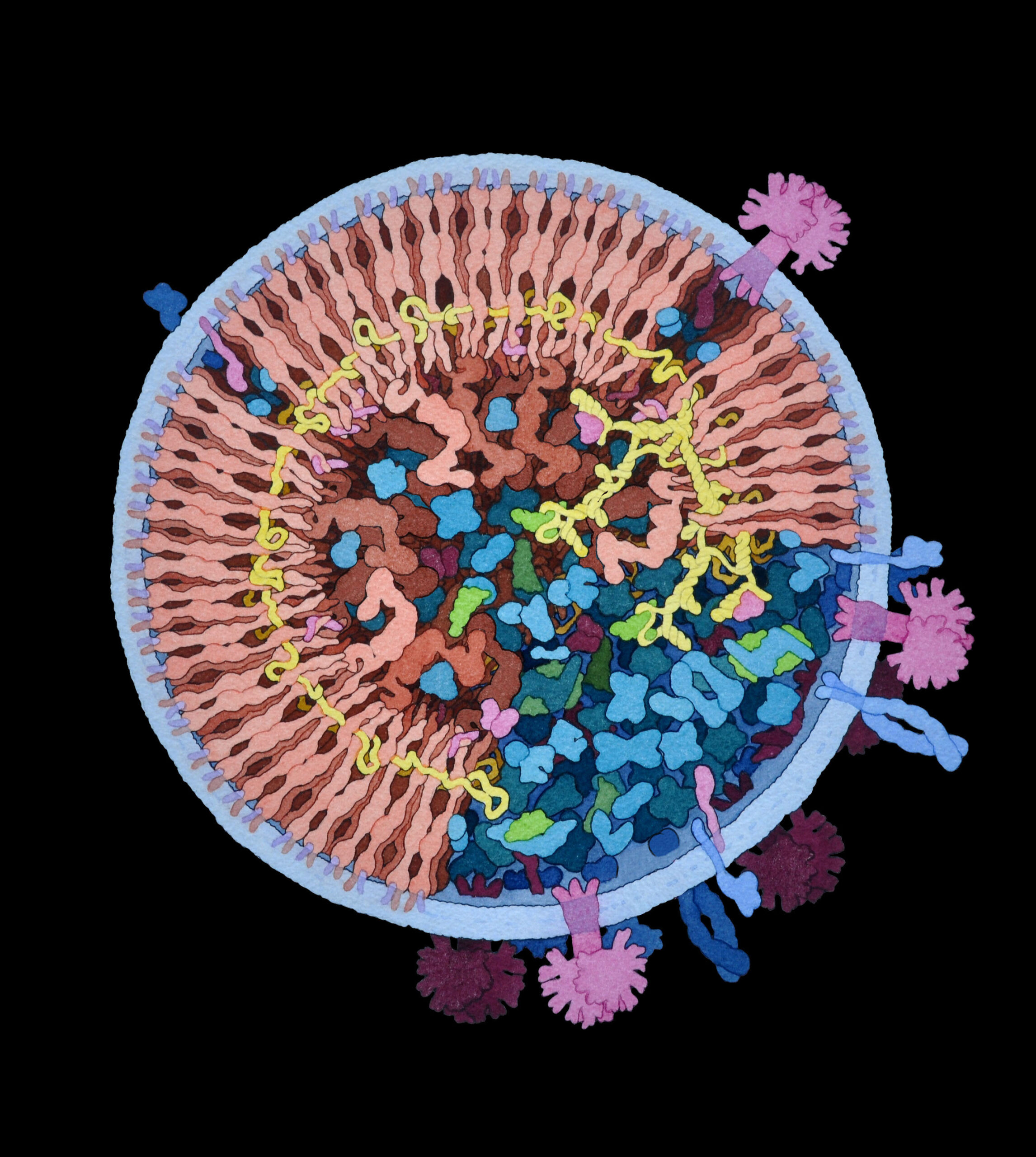

This painting depicts the immature HIV particle, after budding but before maturation. Gag is in light pink, other viral protein is in magenta, RNA is in yellow, cellular protein is in blue and tRNA is in green.
Click here for a description of the scientific references used for the illustration.
Click on the image for a full-size file.
Cross-section of mature HIV (2011, David S. Goodsell)
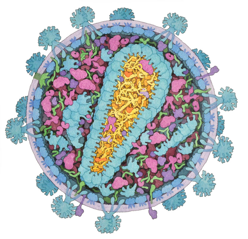
This painting depicts the mature HIV particle, with structural proteins in blue, viral enzymes in magenta, accessory proteins in green and viral RNA in yellow. Host proteins and tRNA are shown in purple.
Several educational resources are available at the Protein Data Bank, using this image in an interactive Flash activity and a poster.
The science behind the painting is described in: BAMBED 40, 291-296 (2012)
Click on the image for a full-size version.