B-HIVE Laboratory Highlight: HIV-1 uncoating location dictates sites of integration
Following the capsid’s trip inside a cell
Once HIV-1 gets inside a cell, the virus faces a delivery problem. Its genetic material is sealed inside the capsid, a cone-shaped protein shell which has to break apart before the viral DNA can be integrated into one of the host cell’s chromosomes. Pinpointing where capsid breakage and integration occur in living cells has been a difficult challenge.
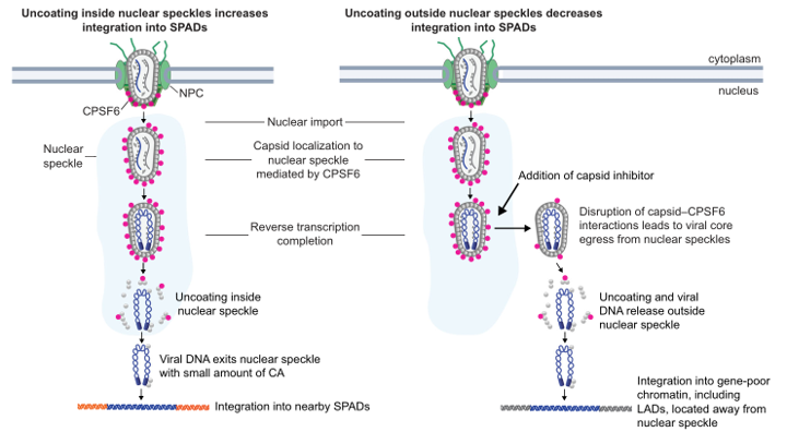
Working at the National Cancer Institute, B-HIVE investigators Ryan Burdick, Vinay Pathak, Wei-Shau Hu and other colleagues have now followed individual viral cores through this final step in cells. They found that the capsid comes apart inside nuclear speckles, and the viral DNA then travels only a very short distance before integrating into nearby chromatin.
Nuclear speckles are droplet-like compartments inside the nucleus. These droplets are made up of a dense collection of RNA-processing proteins that gather near genes that are actively being read. HIV-1 cores are drawn to them with help from a host protein called CPSF6, which binds to the surface of the capsid. The chromatin bordering these speckles is gene-rich and transcriptionally active, characteristic of genomic locations that HIV-1 prefers to incorporate.
Watching a shell break open
To see uncoating directly, the team built virus particles carrying two fluorescent tags at once: a green marker fused to the capsid shell, and a far-red marker floating in the interior of the capsid. Losing the interior signal means the shell has cracked open; losing the green signal means the shell has fallen apart. Speckles were labeled in a separate color in the target cells, so all three could be tracked together under a spinning-disk confocal microscope, frame by frame, about ten hours into infection.
Of the cores the team watched break open, every one lost its interior contents while sitting inside a speckle, and most then lost the shell itself in the same spot. The two events came in quick succession, roughly eight minutes apart, suggesting the capsid shell falls apart soon after the first breach.
Drugs can change where insertions occur
The capsid is a drug target of PF74 (a laboratory compound) and lenacapavir (an FDA approved drug). Both bind the same pocket on the capsid surface that CPSF6 uses. When the team added either drug after the cores had already settled into speckles, the cores were pushed out. CPSF6 fell off within minutes – faster with PF74 than with lenacapavir – and once most of it was gone, the cores drifted out of the speckle and uncoated somewhere else. While PF74 allowed cores to leave speckles largely intact, lenacapavir cracked the cores first and the broken cores lingered before exiting.
That relocation had consequences. Using a fluorescent reporter that lights up actively transcribing proviruses, the team found that proviruses formed after drug-induced uncoating ended up integrating about twice as far from the nearest speckle as untreated ones. Sequencing the integration sites showed that integration into speckle-associated domains dropped sharply, while insertions into gene-poor, lamina-associated domains rose.
What this tells us about HIV-1
The picture that emerges is a tight spatial chain of events: CPSF6 holds the core in a speckle where the capsid shell breaks open, viral DNA exits the capsid, and travels only a short distance to its integration site. The location of uncoating, not just the timing, helps determine which part of the genome will become the site of viral integration. Where a provirus lands determines how actively it is transcribed, and that in turn shapes the latent reservoir that makes HIV-1 so difficult to clear.
Several questions still remain: the researchers note that a small amount of capsid protein stays attached to the viral DNA as it leaves the speckle, and no one yet knows whether that remnant does anything useful. Nor is it clear when the integration machinery assembles, or what guides it to a specific stretch of chromatin once it arrives.
Meet the Researcher
How did you get interested in science?
I became interested in science at a young age, when my father taught my older brother and me to scuba dive in Lake Erie. Exploring the underwater environment sparked a fascination with nature and a curiosity about how the natural world works. That early interest led me to earn a bachelor’s degree in biology and a master’s degree in environmental studies. During graduate school, I worked as an aquatic ecologist for an environmental consulting firm, while my brother pursued a career as a marine biologist specializing in coral reef ecology. After completing my master’s degree, I switched focus and joined the USDA’s National Center for Agricultural Utilization Research, where I studied fungal enzymes with the potential to convert agricultural materials into biofuels. My four years at the USDA deepened my interest in molecular biology and microbiology and ultimately inspired me to pursue biomedical research. In 2008, I joined Vinay Pathak’s laboratory in the HIV Drug Resistance Program—now the HIV Dynamics and Replication Program—at the National Cancer Institute.
Tell us about the lab where you did this work.
This work was conducted primarily in Vinay Pathak’s laboratory, the Viral Mutation Section, within the HIV Dynamics and Replication Program at the National Cancer Institute in Frederick, Maryland. Although I performed most of the experiments, several colleagues made important contributions. Krista Delviks-Frankenberry and Rokeya Siddiqui generated the CPSF6-knockout HeLa cell line, which was particularly challenging to produce. Sean Patro and Xiaolin Wu at the Frederick National Laboratory, together with Stephen Hughes, scientist emeritus in the HIV Dynamics and Replication Program, performed the integration site analysis. This part of the study was essential because it allowed us to connect our microscopy observations with changes in where HIV integrates into the genome. Overall, the study reflects the highly collaborative environment in the Pathak laboratory and across the broader research community in Frederick.
What were the biggest challenges with this study?
The primary challenge was optimizing the live-cell imaging conditions to reliably capture the biological processes we wanted to observe while minimizing photobleaching and phototoxicity. This required carefully balancing signal intensity, temporal resolution, and cell health, because improving one parameter often compromised the others. I systematically tested different combinations of imaging settings until I identified conditions that produced clear images at the required time points while preserving cell viability and signal stability.
What are you working on now?
I’m involved in several projects in the Pathak laboratory. One project builds on our recent work and aims to develop a more detailed mechanistic understanding of HIV-1 uncoating and the events that occur between uncoating and integration. I’m also collaborating with colleagues in the Pathak laboratory to investigate how interactions between host nucleoporins and the capsid surface facilitate passage through the nuclear pore complex, as well as how capsid-targeting inhibitors, such as lenacapavir, block infection. In addition, we are also working with laboratories within and outside the NIH to use our imaging assays to study other aspects of HIV-1 infection.